


Respect the Radiation. Understand the Dose. Keep the Risk in Perspective.
Mammography uses ionizing X-rays, so the radiation exposure is real. But the dose from a standard screening mammogram is low, and for most average-risk women in the recommended screening age range, the benefit of earlier breast-cancer detection outweighs the radiation risk.
“Radiation” is one of those words that immediately sounds dangerous.
And biologically, ionizing radiation absolutely deserves respect.
But radiation risk is not binary.
It depends on:
- dose
- tissue exposed
- age at exposure
- frequency
- the reason the exposure is occurring
A mammogram and an abdominal CT both involve X-rays.
That does not mean they expose the body to the same amount of radiation.
What Makes Mammography Radiation “Ionizing”?
Mammography uses X-rays.
X-rays have enough energy to ionize atoms and molecules.
At the cellular level, ionizing radiation can damage DNA either:
- directly through energy deposition
- or indirectly through reactive chemical species generated in tissue
Cells have extensive repair systems that handle DNA damage constantly.
But no ionizing-radiation exposure should be described as literally risk-free.
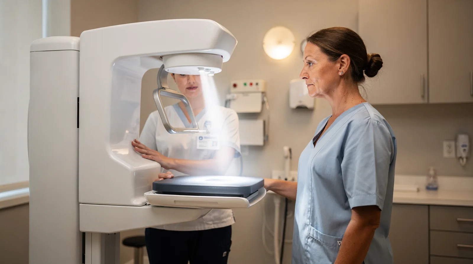
Ionizing Radiation Can Damage DNA. Dose Determines How Much That Matters Clinically.
The existence of a molecular mechanism does not mean every exposure creates a measurable health outcome. Risk depends heavily on dose, age, tissue sensitivity, and cumulative exposure.
How Much Radiation Is in a Mammogram?
A commonly cited effective dose for a standard screening mammogram is approximately:
~0.4 millisieverts (mSv)
That number is an estimate, not a universal patient-specific dose.
Actual exposure varies with:
- breast size and composition
- number of images required
- imaging equipment
- technique
- whether additional diagnostic views are needed
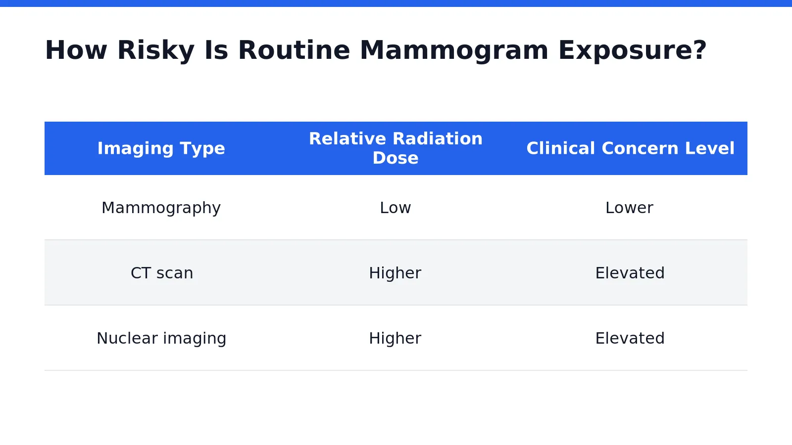
| Imaging Procedure | Typical Effective Dose | Context |
|---|---|---|
| Dental X-ray | ~0.005–0.01 mSv | Very low dose |
| Chest X-ray | ~0.02 mSv | Low dose |
| Mammography | ~0.4 mSv | Low-dose breast imaging |
| CT | ~2–16 mSv depending on exam | Generally much higher than mammography |
| Nuclear Medicine | Wide range | Depends heavily on radiopharmaceutical and study |
Comparing Modalities Is Useful — But Don't Turn the Numbers Into False Precision.
Effective-dose estimates are population-level comparison tools. The exact dose to an individual patient varies, especially when additional views or diagnostic imaging are needed.
Should Mammogram Radiation Worry You?
It should be understood.
It generally should not be feared.
The FDA states that radiation risk from mammography is very small compared with the benefit of detecting breast cancer early.
Current U.S. Preventive Services Task Force guidance recommends:
Screening mammography every 2 years from age 40 through age 74 for average-risk women.
Low Radiation Risk Does Not Mean “No Risk.” It Means the Benefit Generally Wins.
Screening decisions should balance the small radiation exposure and other potential harms of screening against the benefit of finding breast cancer earlier.
Radiation Is Not the Only Mammography Risk Worth Discussing.
This is an important point.
Screening has other potential harms, including:
- false-positive results
- additional imaging
- biopsies that ultimately prove benign
- anxiety
- overdiagnosis and overtreatment
- false-negative results
Those issues belong in an informed screening conversation too.
Does Cumulative Radiation Exposure Matter?
Yes.
Repeated ionizing-radiation exposures contribute to lifetime radiation dose.
But “cumulative” does not mean every X-ray should simply be added into one scary number without context.
Different tissues receive different doses, and age at exposure matters.
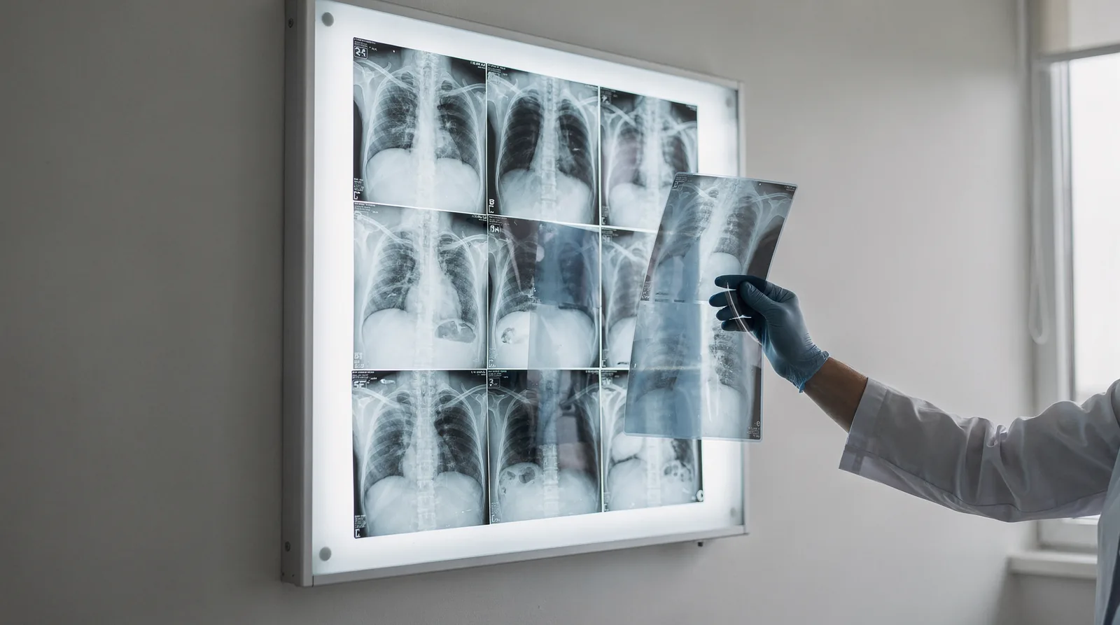
Avoid Unnecessary Radiation. Don't Avoid Necessary Imaging.
The objective is not zero medical radiation at all costs. The objective is appropriate imaging, appropriate dose, and avoidance of unnecessary repeat testing.
Why Is CT Usually a Bigger Radiation Conversation?
CT imaging can deliver substantially more radiation than conventional projection X-rays or mammography.
That does not make CT “bad.”
CT can:
- identify life-threatening bleeding
- diagnose pulmonary embolism
- characterize tumors
- evaluate trauma
- prevent unnecessary exploratory procedures
The dose is justified when the expected diagnostic value outweighs the risk.
Higher Dose Does Not Automatically Mean Wrong Test.
A medically necessary higher-dose scan can provide far more benefit than avoiding radiation while missing an important diagnosis.
Mammography and Nuclear Medicine Are Not the Same Type of Exposure.
Mammography uses an external X-ray source.
Nuclear medicine typically involves administering a radiopharmaceutical that emits radiation from inside the body while a detector measures its distribution.
Dose varies dramatically depending on the study and radionuclide.
This distinction matters because saying simply:
“I had radiation imaging”
tells us almost nothing about actual dose or risk.
Radiation Conversations Need Dose, Route, Isotope or Modality, and Tissue Context.
That is the same discipline used in nuclear pharmacy and medical imaging: identify the radiation source, understand where it goes, quantify the dose, and interpret the exposure in the context of the clinical benefit.
Can You “Detox” Radiation After a Mammogram?
No.
Once the X-ray exposure is over, there is no radiation left inside the body from the mammogram.
Mammography does not make the patient radioactive.
There is nothing sitting in the breast afterward that needs to be “flushed out.”
X-Ray Exposure Is an Energy Event — Not a Toxin Left Behind.
Mammography exposes tissue to X-ray photons for a brief period. When the X-rays stop, the exposure stops. Supplements, juices, saunas, charcoal, or “detox” products cannot remove radiation that is no longer present.
What About Antioxidants After Radiation Exposure?
This is where biology and supplement marketing often get mixed together.
Ionizing radiation can generate reactive species.
The body already has endogenous antioxidant and repair systems, including:
- glutathione systems
- superoxide dismutase
- catalase
- DNA-repair pathways
Nutrients participate in normal cellular metabolism and antioxidant enzyme function.
But that does not establish that taking an antioxidant supplement around a mammogram reduces future cancer risk.
Mechanism Is Not the Same as a Proven Protective Intervention.
“This nutrient participates in antioxidant biology” does not prove that taking more of it before or after mammography prevents radiation-induced DNA damage or cancer.
Where Does Core 3 Fit?
As general nutritional support.
Not as radiation protection.
Core 3 Foundation
Core 3 combines a multivitamin, omega-3 fish oil, and probiotic/prebiotic component into one simplified AM routine.
Those categories can support broad nutritional adequacy and general health. Core 3 should not be marketed as preventing mammography radiation damage or as a post-imaging detox.
Explore Core 3 Foundation →What About Complete Core 5?
Same principle.
Complete Core 5 provides broader nutritional coverage by adding PM magnesium + zinc to Core 3.
Magnesium and zinc participate in many normal enzymatic processes.
That still does not make Core 5 an anti-radiation product.
Complete Core 5 Foundation
Core 3 in the morning plus PM Magnesium + Zinc for adults who independently want that broader nutritional foundation.
Use it because the nutritional categories fit the person — not because they had an X-ray.
Explore Complete Core 5 →What Actually Supports Long-Term Cellular Health?
The boring answers remain the strongest:
- do not smoke
- maintain appropriate body weight
- eat a nutrient-dense diet
- exercise regularly
- sleep adequately
- limit unnecessary medical radiation
- use necessary imaging when the clinical benefit justifies it
- participate in appropriate cancer screening
Nutrition Supports the Person. It Does Not Cancel the Radiation Dose.
Good nutrition matters every day. That is fundamentally different from claiming a particular supplement neutralizes the biological effect of an X-ray exposure.
Should You Track Every Radiation Exposure?
Keeping an accurate medical-imaging history can be useful.
But patients should not feel responsible for calculating an exact lifetime cancer-risk score from every dental X-ray, mammogram, CT, and nuclear medicine study.
More useful questions include:
- Is this imaging medically necessary?
- Is there a prior study that already answers the question?
- Could ultrasound or MRI answer the question without ionizing radiation?
- Will the result change management?
- Is the facility using appropriate dose-optimization standards?
Appropriate Imaging + Dose Optimization Beats Fear.
Avoid duplicate or unnecessary imaging when possible. But do not decline a clinically important study simply because it involves radiation.
When Should Screening Mammography Start?
For average-risk women, the U.S. Preventive Services Task Force currently recommends:
Mammography every 2 years from ages 40 through 74.
That recommendation does not cover every possible individual situation.
Screening plans may differ for people with:
- BRCA1 or BRCA2 variants
- other high-risk genetic syndromes
- strong family history
- prior breast cancer
- high-risk breast lesions
- high-dose chest radiation at a young age
Average Risk and High Risk Are Different Screening Conversations.
People at elevated breast-cancer risk may need earlier, more frequent, or different imaging. That plan should be individualized with the appropriate clinician.
What About Dense Breasts?
Dense breast tissue can make mammography more difficult to interpret and is itself associated with higher breast-cancer risk.
U.S. mammography facilities now provide breast-density information in patient reports.
But whether every person with dense breasts should automatically receive supplemental ultrasound or MRI remains less settled.
The USPSTF currently considers the evidence insufficient to determine the overall balance of benefits and harms of routine supplemental ultrasound or MRI after a negative mammogram solely because of breast density.
Five Mammography Radiation Myths Worth Killing
False. Mammography uses external X-rays. Once the exposure ends, the patient is not radioactive.
False. Both use ionizing X-rays, but typical CT doses are usually substantially higher.
False for most average-risk women in the recommended screening age range. Risk and benefit depend on dose and clinical context.
Unsupported. No supplement has been established as a routine post-mammogram radiation antidote.
Also false. Ionizing radiation carries a small potential carcinogenic risk. Screening is used because the expected benefit exceeds that small risk.
Don't Minimize Radiation. Don't Weaponize It Either.
Mammography uses real ionizing radiation. The dose is low. The risk is small. The screening benefit is meaningful for appropriate patients. That entire sentence can be true at the same time.
Frequently Asked Questions
Is mammogram radiation dangerous?
Mammography uses ionizing radiation, so the risk is not literally zero. However, the dose from routine screening is low, and for most people in recommended screening populations, the benefit of detecting breast cancer earlier outweighs the small radiation risk.
How much radiation is in a mammogram?
A commonly cited effective dose for standard mammography is about 0.4 mSv, although actual patient exposure varies with equipment, breast characteristics, number of views, and whether diagnostic imaging is required.
Is mammography less radiation than CT?
Generally yes. Typical CT effective doses often fall several-fold to many-fold above standard mammography, although dose depends on the specific CT examination.
Does a mammogram make you radioactive?
No. Mammography uses an external X-ray beam. Nothing radioactive is administered, and no radioactive material remains in the body afterward.
Do I need to detox after a mammogram?
No. There is no radiation substance to remove from the body after an X-ray-based mammogram.
Should I take glutathione or antioxidants after a mammogram?
No routine antioxidant supplement has been established as a post-mammogram intervention that reduces future cancer risk. Maintain good nutrition because it supports overall health, not because it “cancels” the X-ray dose.
Does Core 3 protect against mammogram radiation?
No. Core 3 is a broad nutritional foundation containing a multivitamin, omega-3, and probiotic/prebiotic component. It should not be marketed as radiation protection.
Does Complete Core 5 protect DNA from mammography?
There is no clinical evidence establishing Complete Core 5 as protection against mammography-related DNA damage. Magnesium and zinc participate in normal biological processes, but that is not the same as demonstrating protection from medical radiation.
Should I skip mammography because I have already had many X-rays?
Not automatically. Prior exposure can be discussed with your clinician, but medically appropriate screening should not be abandoned simply because other imaging has occurred.
What is the current screening recommendation?
The U.S. Preventive Services Task Force recommends mammography every two years for average-risk women ages 40 through 74. People at higher-than-average risk may need a different screening plan.
Key Evidence
U.S. Food & Drug Administration — Mammography.
The FDA describes mammography as low-dose X-ray imaging and states that the benefits of early breast-cancer detection outweigh the radiation risk.
FDA Mammography Information →
FDA Medical Imaging Dose Comparison.
FDA reference material lists an average adult effective dose around 0.4 mSv for mammography compared with approximately 2–16 mSv for CT, depending on the examination.
FDA Radiation Dose Comparison →
U.S. Preventive Services Task Force.
Current guidance recommends biennial screening mammography from ages 40 through 74 for average-risk women and concludes that this strategy has a moderate net benefit.
USPSTF Recommendation →
National Cancer Institute.
NCI recognizes radiation exposure as a potential harm of mammography but describes the cancer risk as very low and discusses other screening tradeoffs including false positives, overdiagnosis, and false negatives.
NCI Breast Cancer Screening →
Respect the Radiation. Understand the Dose. Don't Turn Screening Into Fear.
Mammography uses ionizing radiation. That exposure carries a small risk. The dose is low, the screening benefit can be substantial, and no supplement has been established as a way to neutralize the exposure afterward.
The Takeaway
Mammography uses real ionizing X-rays.
The dose from routine screening is low.
A commonly cited effective dose is approximately 0.4 mSv.
CT examinations generally involve considerably more radiation.
Screening mammography does not leave radioactive material inside the body.
There is no evidence-based “radiation detox” required after a mammogram.
Antioxidant biology is real, but that does not prove supplements prevent mammography-related cancer risk.
Core 3 and Complete Core 5 can support general nutritional adequacy, but they are not radiation-protection products.
Avoid unnecessary radiation when practical.
Do not avoid medically appropriate imaging because of exaggerated radiation fear.
Know the modality. Know the dose. Know the benefit. Then make the decision.
This article is for educational purposes only and is not medical advice. Breast-cancer screening decisions should be individualized according to age, breast-cancer risk, family history, genetic risk, prior breast disease, breast density, previous radiation exposure, symptoms, and clinician guidance. Do not delay or decline medically appropriate breast imaging solely because of information in this article. Dietary supplements have not been established as a method for preventing or reversing radiation-related effects from mammography.


